Flow-Cytometric Work-Up in Chronic Lymphoproliferative Disorders
๐ 10 slides
๐ 58 views
๐
1/23/2026
Introduction to CLPDs
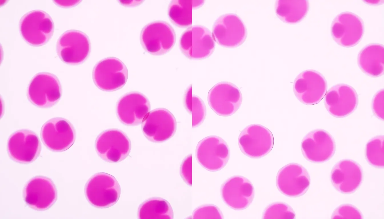
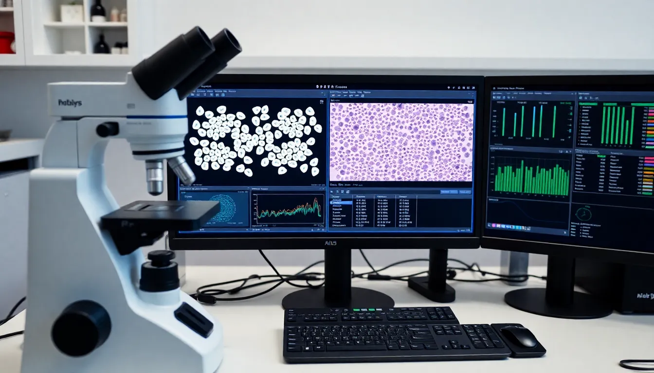
Chronic Lymphoproliferative Disorders (CLPDs) are clonal lymphoid proliferations with indolent behavior.
2
Indications for Flow Cytometry
- Persistent lymphocytosis (>5,000/ยตL) lasting >3 months.
- Atypical lymphoid cells in peripheral blood or bone marrow.
- Essential for staging and minimal residual disease assessment.
3
Specimen Handling
- Optimal specimens: peripheral blood (EDTA), bone marrow, or tissue suspensions.
- Anticoagulants affect viability; process within 24-48 hours.
- Cell viability >70% required for reliable results.
4
Flow Cytometry Principles
- CD45/SSC gating strategy to identify lymphoid populations.
- Light scatter distinguishes small vs. large lymphoid cells.
- Internal controls (normal B/T cells) validate antibody performance.
5
Core Antibody Panel
- B-cell: CD19, CD20, CD22, CD79b, ฮบ/ฮป light chains.
- T-cell: CD3, CD5, CD7, CD2, CD4/CD8 ratio.
- NK-cell: CD16, CD56, CD57.
6
B-Cell CLPD Signatures
- CLL: CD5+, CD23+, CD200+, CD43+, weak surface Ig.
- MCL: CD5+, CD23-, FMC7+, Cyclin D1+ (by IHC).
- HCL: CD11c+, CD25+, CD103+, CD123+.
7
T/NK-Cell CLPDs
- T-LGL leukemia: CD3+, CD8+, CD57+, TCRฮฑฮฒ+.
- Sรฉzary syndrome: CD4+, CD7-, CD26- circulating cells.
- NK-LGL leukemia: CD3-, CD56+, CD16+, cytotoxic markers+.
8
Diagnostic Pitfalls
- Reactive lymphocytosis may mimic CLPD immunophenotype.
- Antigen modulation after therapy causes false negatives.
- Always correlate with morphology and clinical history.
9
Multimodal Integration
- Morphology confirms atypical cells seen by flow.
- Cytogenetics (e.g., t(11;14) in MCL) adds specificity.
- Molecular studies (IGH rearrangement) confirm clonality.
10
Key Takeaways
- Flow cytometry is the cornerstone of CLPD diagnosis.
- Standardized panels reduce diagnostic errors.
- WHO classification integrates flow with other modalities.
1 / 10