Liver Cancer Detection Using Image Processing

📑 5 slides 👁 6 views 📅 3/1/2026 📄 PDF
0.0 (0 ratings)

Introduction to Liver Cancer Detection

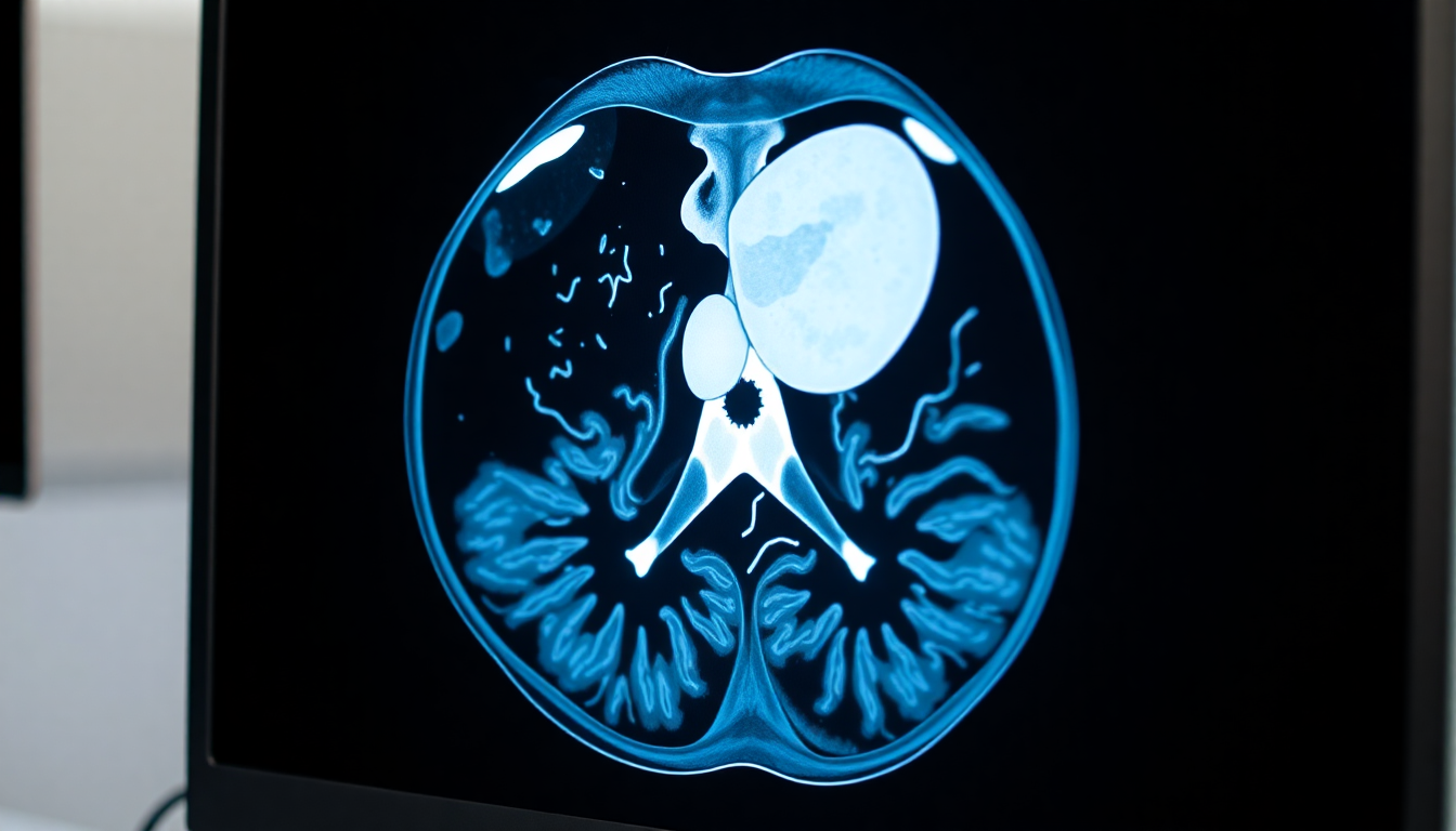
Liver cancer survival rate in India is only 5%, highlighting the need for early detection.

Introduction to Liver Cancer Detection
2

Image Processing Techniques

  • Preprocessing involves noise reduction using median and anisotropic diffusion filtering.
  • Histogram enhancement improves image contrast for better analysis.
  • Segmentation uses fuzzy centroid-based region-growing model for tumor detection.
  • Adaptive thresholding helps in partitioning the liver accurately.
Image Processing Techniques
3

Literature Review Highlights

  • Multiresolution Fractal (MF) feature achieves 90% accuracy in liver disease identification.
  • Functional Link Neural Network shows high diagnostic rates for liver diseases.
  • Fuzzy descriptors and rules improve liver region identification in CT scans.
  • Wavelet transform features offer 90% classification accuracy for liver tissue.
Literature Review Highlights
4

Prior Work and Innovations

  • Connected compounds numbering algorithm (CCL) automates liver area extraction.
  • Neuro-fuzzy systems combine fuzzy logic and neural networks for tumor detection.
  • Watershed and Ostu's methods enhance MRI images for cancer cell separation.
  • SVM classifier improves tumor segmentation accuracy in CT scans.
Prior Work and Innovations
5

Conclusion and Future Directions

  • Proposed method effectively segments tumors with radiologist-validated results.
  • Future work includes expanding feature vectors for other liver disease classifications.
  • Technique enhances, segments, and extracts liver areas with high accuracy.
  • Potential to improve diagnostic training for new medical professionals.
Conclusion and Future Directions
1 / 5